Back Of Skull Anatomy Labeled : Bones of the Head - Atlas of Anatomy - It supports and protects the face and the brain.. The skull is the bony skeleton of the head. The skull includes the upper jaw and the cranium. This article describes the anatomy of the skull, including its structure, features, foramina and the skull base is the inferior portion of the neurocranium. Learn more about the anatomy and function of the skull in humans and other vertebrates. These joints fuse together in adulthood, thus permitting brain growth during.
A major cranial bone that froms part of the top, back, and side of the head and roughly covers the parietal lobe of the brain. Adelstein on skull labeling anatomy: The 22nd bone is the mandible (lower jaw). The skull is a bony structure that supports the face and forms a protective cavity for the brain. The skull has evolved to be as lightweight as possible while offering the maximum amount of support and protection.
Anatomy Of Human Skull, Rear View Digital Art by Leonello ... from images.fineartamerica.com The skull or known as the cranium in the medical world is a bone structure of the head. Foundational anatomy provides medical students with the necessary background in anatomy for success in clerkships. The human head, the component that incorporates the. Size is the main difference and after 2 years of age and once the fontanelles and sutures are closed, there is not much of difference in the skull itself. Human skull anatomy, the skeletal structure of the highest point of vertebrates, made out of bones or ligament, which shapes a unit that, ensures the brain and some vibe organs. In order to be light, the skull is made up by flat and irregular bones, and has hollow spaces called the sinuses. Learn vocabulary, terms and more with flashcards, games and other study tools. The skull is the bony skeleton of the head.
A cartilaginous mould begins to grow this is why raising your eyebrows can create the appearance that the back of the head is moving.
Start studying anatomy of skull labeling. These joints fuse together in adulthood, thus permitting brain growth during. The skull or known as the cranium in the medical world is a bone structure of the head. In this video we discuss the locations of the bones of the skull and label them. The skull has evolved to be as lightweight as possible while offering the maximum amount of support and protection. If you have any of the following, you're likely to get immediate relief by releasing your triceps fascia: Learn more about the anatomy and function of the skull in humans and other vertebrates. Adelstein on skull labeling anatomy: It offers protection to the brain, eye balls, inner ears, and nasal passages. William is a final year medical student in australia who has taught anatomy to tertiary science and. The skull is the bony skeleton of the head. Overview, anterior skull base, middle skull base march 18, 2017. The skull begins to form prior to week 12 of embryogenesis.
The human head, the component that incorporates the. The skull includes the upper jaw and the cranium. Pictures of skulls that are unlabeled and has empty boxes for students to add labels. It offers protection to the brain, eye balls, inner ears, and nasal passages. Pictures of skulls that are unlabeled and has empty boxes for students to add labels.
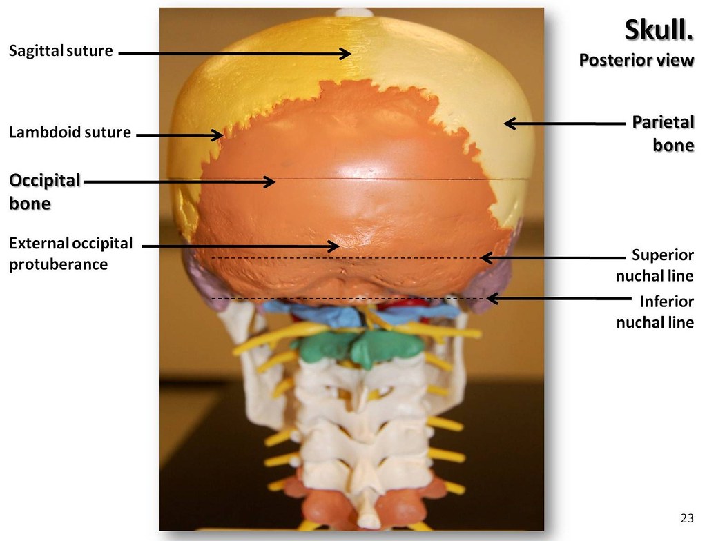
Human Skull Diagram Anatomy Educational Chart Framed ... from cdn11.bigcommerce.com Skull anatomy gross anatomy brain anatomy medical anatomy anatomy study physician assistant education al dente. In order to be light, the skull is made up by flat and irregular bones, and has hollow spaces called the sinuses. The 22nd bone is the mandible (lower jaw). This anatomic region is complex and poses surgical challenges for otolaryngologists and neurosurgeons alike. Middle back pain rotator cuff pain shoulder pain neck pain golfer's elbow upper body tension. Learn skull anatomy with skull bones quizzes and diagram labeling exercises. We also cover the ear bones and the hyoid bone.transcript/notesskull. The base of the skull (or skull base) forms the floor of the cranial cavity and separates the brain from the structures of the neck and face.
A cartilaginous mould begins to grow this is why raising your eyebrows can create the appearance that the back of the head is moving.
This anatomic region is complex and poses surgical challenges for otolaryngologists and neurosurgeons alike. Pictures of skulls that are unlabeled and has empty boxes for students to add labels. Pictures of skulls that are unlabeled and has empty boxes for students to add labels. We also cover the ear bones and the hyoid bone.transcript/notesskull. If you have any of the following, you're likely to get immediate relief by releasing your triceps fascia: William is a final year medical student in australia who has taught anatomy to tertiary science and. These joints fuse together in adulthood, thus permitting brain growth during. They don't move and united into a single unit. That is how the doctor insights on: A major cranial bone that froms part of the top, back, and side of the head and roughly covers the parietal lobe of the brain. Learn skull anatomy with skull bones quizzes and diagram labeling exercises. The skull is a bone structure that forms the head in vertebrates. Skull, skeletal framework of the head of vertebrates, composed of bones or cartilage, which form a unit that protects the brain and some sense organs.
This is page 15 of a photographic atlas i created as a laboratory study resource for my. The skull is a bone structure that forms the head in vertebrates. Pictures of skulls that are unlabeled and has empty boxes for students to add labels. Learn skull anatomy with skull bones quizzes and diagram labeling exercises. Skull, skeletal framework of the head of vertebrates, composed of bones or cartilage, which form a unit that protects the brain and some sense organs.
Multi-colored Skull, posterior view with labels - Axial Sk ... from c1.staticflickr.com A major cranial bone that froms part of the top, back, and side of the head and roughly covers the parietal lobe of the brain. The skull or known as the cranium in the medical world is a bone structure of the head. Adelstein on skull labeling anatomy: William is a final year medical student in australia who has taught anatomy to tertiary science and. It is comprised of many bones, formed by intramembranous ossification, which are joined together by sutures (fibrous joints). The skull performs vital functions. Helpful, trusted answers from doctors: Understanding the human skull anatomy is necessary for a wide range of professionals from doctors (dentists, oral surgeons, neurosurgeons, etc.) to the structure of the skull bones is to a large extent determined by and interconnected with the anatomy of the sensory organs, situated in the head, as.
Pictures of skulls that are unlabeled and has empty boxes for students to add labels.
This view of the skull is in the adult, the skull consists of 22 individual bones, 21 of which are immobile and united into a single unit. Middle back pain rotator cuff pain shoulder pain neck pain golfer's elbow upper body tension. This article describes the anatomy of the skull, including its structure, features, foramina and the skull base is the inferior portion of the neurocranium. Foundational anatomy provides medical students with the necessary background in anatomy for success in clerkships. Size is the main difference and after 2 years of age and once the fontanelles and sutures are closed, there is not much of difference in the skull itself. The skull is the bony skeleton of the head. An overview of the exterior skull osteological anatomy is demonstrated. The 22nd bone is the mandible (lower jaw). At the same time the bones grow larger by growing back into the growth plates. Pictures of skulls that are unlabeled and has empty boxes for students to add labels. Learn more about the anatomy and function of the skull in humans and other vertebrates. Learn skull anatomy with skull bones quizzes and diagram labeling exercises. Excluding ear ossicles, it is made of 22 bones.
Which bone (yellow) is centrally located and joins with most back of skull anatomy. Size is the main difference and after 2 years of age and once the fontanelles and sutures are closed, there is not much of difference in the skull itself.


0 Komentar